The short answer
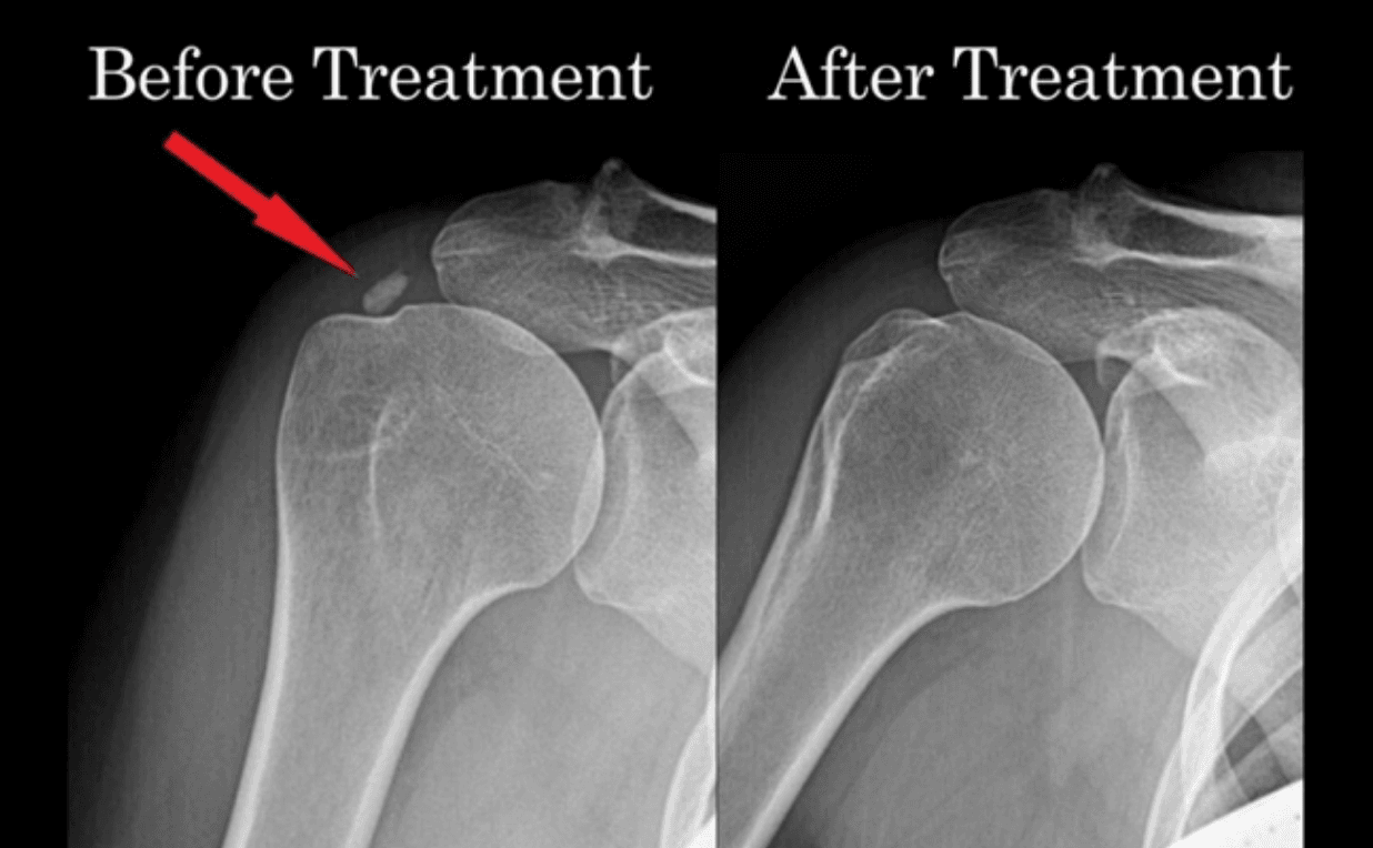
Calcific tendonitis is a buildup of calcium (hydroxyapatite) within the rotator cuff. Because there is only about 8mm of space between the shoulder bones, the deposit causes inflammation and pain, especially with overhead motion. Treatment includes physical therapy, cortisone, and ultrasound-guided barbotage/percutaneous tenotomy (Tenex) to break up and remove the calcium.
Medically reviewed by Marc Gruner, DO, MBA, RMSK
Sports medicine physician, Mayo Clinic fellowship. Last reviewed June 2026
Overview
Calcific tendonitis develops when calcium — in the form of hydroxyapatite — accumulates within the rotator cuff tendons. With only about 8 millimeters of space between the shoulder bones, the deposit crowds the joint, causing inflammation and pain that is often worst with overhead movement and can make physical therapy difficult.
When conservative care isn't enough, Dr. Gruner can visualize the calcium under ultrasound and guide a needle to break up and aspirate it (a percutaneous, Tenex-style procedure), relieving pain while avoiding open surgery, which is reserved as a last resort.
Common symptoms
- Pain with shoulder movement, especially overhead
- Difficulty completing physical therapy due to pain
What to expect at your visit
- 1.
A focused conversation
Dr. Gruner reviews your history, prior imaging, and goals — what you want to get back to doing.
- 2.
Point-of-care ultrasound
When useful, a diagnostic musculoskeletal ultrasound evaluates the injury in real time, with no radiation.
- 3.
A clear, individualized plan
You leave understanding the diagnosis, the non-surgical options, and the expected timeline — in plain language.
Rehabilitation protocol
What to expect week by week after your procedure. Your plan is individualized; follow these alongside your physical therapist.
Shoulder — Ultrasound-Guided Lipogems / PRP Protocol
Week 1
Protect, reduce pain and swelling
- Sling for 7 days; begin weaning after day 3
- No lifting > 5 lbs; avoid overhead activity 2 weeks
- Don't sleep on the procedure side
- Begin gentle active ROM (pendulums) 4×/day
Week 2
Begin active range of motion
- No lifting > 10 lbs; avoid overhead activity
- Continue AROM and PROM
- Begin isometrics and thoracic mobility
- Stop any exercise if pain > 4/10
Weeks 3–6
Shoulder strengthening
- May lift 10–20 lbs; avoid eccentrics
- Continue active ROM
- Progressive strengthening
Weeks 6–12
Return to sport
- Activities as tolerated
- Begin eccentric training/loading
- Sport-specific exercises
Do not use NSAIDs (ibuprofen, Aleve, aspirin) for 4 weeks, and avoid ice for the first week — both blunt the healing response. Acetaminophen (Tylenol) is fine for discomfort. Care is tailored to you; use these guidelines with your physical therapist.
Frequently asked questions
- How is calcific tendonitis treated without surgery?
- First-line care is physical therapy and a cortisone injection. For persistent deposits, an ultrasound-guided percutaneous procedure (Tenex/barbotage) can break up and remove the calcium at the point of care, avoiding open surgery.