The short answer
Knee arthritis is the wearing of cartilage between the femur and tibia, causing pain and swelling. Non-surgical options include activity modification, weight management, physical therapy, cortisone or gel (viscosupplementation) injections, and orthobiologics such as PRP and microfragmented fat (Lipogems). Knee replacement is reserved as a last resort.

Medically reviewed by Marc Gruner, DO, MBA, RMSK
Sports medicine physician, Mayo Clinic fellowship. Last reviewed June 2026
Overview
Knee osteoarthritis develops as the smooth cartilage lining the joint between the thigh bone (femur) and shin bone (tibia) breaks down, causing pain, swelling, and stiffness. It commonly follows prior trauma, the natural shape of the knee, meniscus injury, and normal aging.
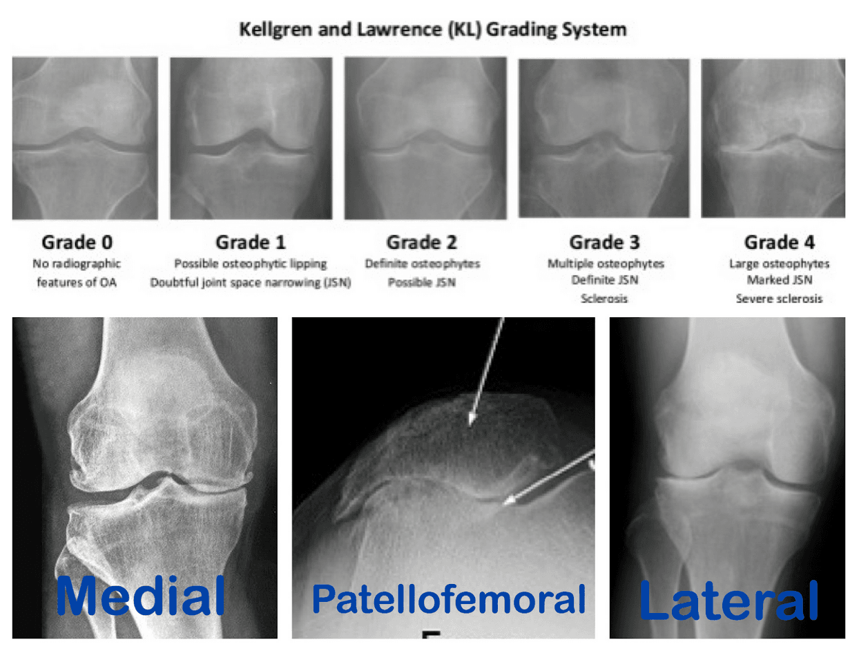
Pain may be felt on the inside (medial) or outside (lateral) of the knee, or in the front from patellar arthritis. Dr. Gruner builds a plan that starts conservative — activity change, weight strategy, and physical therapy — and adds image-guided injections or orthobiologics (PRP, microfragmented fat) for the right candidate, aiming to delay or avoid knee replacement. Cortisone relief tends to be short term, and it should not be repeated more than three times in a year.
Common symptoms
- Pain on the inside or outside of the knee
- Pain in the front of the knee (patellar arthritis)
- Joint swelling after activity
- Stiffness and reduced range of motion
Videos from Dr. Gruner
Orthobiologics for Knee Pain — Dr. Marc Gruner
Is PRP the Answer to Chronic Knee Pain? — Dr. Marc Gruner
Knee Osteoarthritis: More Common Than You Think — Dr. Marc Gruner
Knee Gel Injections for Knee Pain Relief — Dr. Marc Gruner
Knee Pain? Try This Before Surgery — Dr. Marc Gruner
What to expect at your visit
- 1.
A focused conversation
Dr. Gruner reviews your history, prior imaging, and goals — what you want to get back to doing.
- 2.
Point-of-care ultrasound
When useful, a diagnostic musculoskeletal ultrasound evaluates the injury in real time, with no radiation.
- 3.
A clear, individualized plan
You leave understanding the diagnosis, the non-surgical options, and the expected timeline — in plain language.
Rehabilitation protocol
What to expect week by week after your procedure. Your plan is individualized; follow these alongside your physical therapist.
Knee & Hip Joint — Ultrasound-Guided PRP / Adipose / BMAC Injection Protocol
Day 1–2
Protect the joint
- Weight-bear as tolerated; crutches as needed
- Keep bandage dry; no submerging for 72 hours
- Gentle ROM as tolerated; monitor wound
Day 3–7
Begin gentle loading
- Avoid excessive joint loading
- Start low-grade closed-chain program
- Light squats/lunges (bodyweight); optional BFR
Day 8–14
Add resistance
- Avoid impact and heavy lifting to the joint
- Light open-chain exercises; add resistance as tolerated
- May begin swimming and low-resistance biking
Day 14–28
Functional progression
- Avoid impact activities
- Progress functional program; light agility and proprioception
- Resume light aerobic activity (walking)
Beyond 28 days
Long-term prevention
- Establish a long-term home exercise program
- Correct biomechanical issues that contributed to the injury
Do not use NSAIDs (ibuprofen, Aleve, aspirin) for 4 weeks, and avoid ice for the first week — both blunt the healing response. Acetaminophen (Tylenol) is fine for discomfort. Care is tailored to you; use these guidelines with your physical therapist.
Clinical evidence
Selected peer-reviewed research supporting these approaches.
- Platelet-rich plasma versus hyaluronic acid in the treatment of knee osteoarthritis: a meta-analysisPubMed Central PMC7488405
- Platelet-Rich Plasma Versus Hyaluronic Acid for Knee Osteoarthritis: A Systematic Review and Meta-analysis of RCTsPubMed 32302218
- Platelet-Rich Plasma Combined With Hyaluronic Acid Improves Pain and Function Compared With Hyaluronic Acid Alone in Knee OsteoarthritisPubMed 33278533
- Microfragmented Adipose Tissue Injection (MFAT) May Be a Solution to the Rationing of Total Knee Replacement: 2-Year AnalysisPubMed Central PMC8211497
- Two-year clinical outcomes of autologous microfragmented adipose tissue in elderly patients with knee osteoarthritisPubMed 33822274
Frequently asked questions
- Can knee arthritis be treated without surgery?
- Yes. Most patients improve with activity modification, weight management, physical therapy, and injections — including cortisone, viscosupplementation (gel), PRP, or microfragmented fat. Knee replacement is generally a last resort when these measures no longer provide relief.
- Does PRP work for knee arthritis?
- For many patients with mild-to-moderate knee osteoarthritis, ultrasound-guided PRP can reduce pain and improve function, with benefits that studies suggest may last a year or longer. Dr. Gruner reviews your imaging and goals to determine whether you are a candidate.